Wednesday, December 29, 2010

Musculoskeletal Ultrasound Growth Noteworthy

The popularity of musculoskeletal ultrasound (MSK-US) has taken off in recent years with the addition of portable ultrasound technology and greater imaging resolution.  This area of growth has not been fueled by radiologists, instead many physicians in sports medicine, general practice and rheumatology are driving this phenomenon.  I work in the health care educational field, and I have noticed out courses in MSK are very popular.  Our next general course in MSK-US is Feb 26-27 here in Houston.   Please visit www.aheconline.com for more information.  I think MSK-US is a wonderful tool for examining all sorts of aches and pains, and I predict it will continue to grow as more health care providers discover the benefits.  Here is part of an article I found at Auntminnie that explains this topic further.  Cheers!

December 2, 2010 -- CHICAGO - Musculoskeletal ultrasound usage nearly quadrupled between 2000 and 2008, driven primarily by increased utilization by nonradiologists, according to a study presented Thursday at the 2010 RSNA meeting in Chicago.
http://www.auntminnie.com/index.aspx?sec=sup&sub=ult&pag=dis&itemId=93452

Thursday, December 16, 2010

Stress Echocardiography New and Improved?

Some news came out today regarding one of the FDA approved ultrasound contrast agents regarding the use of contrast when performing stress echocardiography. 

No. BILLERICA, Mass. (December 13, 2010)Lantheus Medical Imaging, Inc., a worldwide leader in diagnostic medical imaging, today announced that the U.S. Food and Drug Administration (FDA) has accepted for review the company’s supplemental New Drug Application (sNDA) for DEFINITY® (Perflutren Lipid Microsphere) Injectable Suspension for use in stress echocardiography.  DEFINITY® is currently indicated for use in patients with suboptimal echocardiograms to opacify the left ventricular chamber and to improve the delineation of the left ventricular endocardial border1.  With this acceptance, the FDA will now review the sNDA to determine whether or not to broaden the FDA-approved indication for DEFINITY® to include its use with exercise and pharmacologic stress testing.

Several labs have been using intravenous ultrasound contrast for years when performing stress echos because the border definition is better for most studies when contrast is applied.  One of the downsides to a stress echo is the loss of wall definition at peak exercise even in patients who have fairly good windows.  Contrast adds an extra level of insurance that the walls will be seen better in most echos.  It is news to me that the FDA has not approved contrast for use in stress echo, but has approved it for many years in resting echocardiography.  Perhaps the up side here is that labs can now be re-reimbursed for using contrast in this capacity.  I had erroneously thought that the FDA had already approved intravenous ultrasound contrast for both resting and stress echos.  Live and learn.
http://www.radiopharm.com/News-Press-2010-1213.html

Wednesday, December 15, 2010

Man is Told He is Pregnant in the UK

A man in the UK has been sent a letter by the state run health service informing him that he is pregnant.  Last time I heard of this happened it involved a California Govinator in a Hollywood movie.  I guess this goes in the same category as family pets being sent credit card applications.  I wonder who will host the baby shower.


Hilton Plettell should expect the pitter-patter of tiny feet in seven months’ time, according to the correspondence sent to him from the obstetric department of a hospital in Norwich.
 
The 50-year-old, from Kings Heath, said he was “flabbergasted” when he received the appointment letter inviting him to a scan – and warning him he could be expecting twins!

Opening “Dear Mr Plettell”, the note also advised him: “Please also attend with a full bladder, which is important for your planned ultrasound. An ultrasound scan is used to check the pregnancy, to determine how far pregnant you are, and sometimes it can show twins.”

Hilton, who never got round to having children and works as a merchandising manager, said: “It came as a bit of a shock, I’ve shown the letter to quite a few friends and they can’t believe it.

“I knew the letter was genuine. The date of birth, the National Insurance number and everything on there is correct. I’m just surprised they can send something like that.”


Read More http://www.birminghammail.net/news/birmingham-news/2010/12/08/kings-heath-man-stunned-after-receiving-hospital-letter-telling-him-he-is-pregnant-97319-27786120/#ixzz18CZkghjS


Monday, December 6, 2010

HE is on HIS Way

An ecumenical Christian nonprofit group in England has created a unique Advent advertising campaign. Starting Monday, and running through December 20, billboards and posters will show a mock-up of an ultrasound of baby Jesus, complete with halo. The words are simple: “He’s on His way . . . Christmas starts with Christ.”

Merry Christmas All

http://online.worldmag.com/2010/12/03/ultrasound-of-baby-jesus/

Thursday, October 7, 2010

K-Mart to Screen for Triple A's

Recently, I have been very busy with work, and have not had much time to post.  I came across something on Sonoworld ( www.sonoworld.com ) that is worthy of attention.  It seems many K-marts around the country have begun screening patients in their pharmacy's for abdominal aortic aneurysms (AAA).  They have teamed up with a few good organizations including the SVU to provide this free service.  No where in the information is there a reference to the qualifications of the health care providers being used to screen these patients.  As a Registered Vascular Sonographer, I think it is important to point out to patients that Sonographers are medical professionals, and we specialize in ultrasound.  Many states limit the use of ultrasound in the medical field to those who have demonstrated a proficiency by sitting for written boards, and passing the medical tests needed to gain a credential in medical ultrasound (RVT, or RVS) through either the ARDMS, or CCI.  I am wondering if K-Mart is using registered vascular sonographers?  Any input is welcome.

Kmart Pharmacy Joins Fight Against Abdominal Aortic Aneurysms (AAA)

Retail Partner for Find the AAAnswers Campaign Will Support Free Education, Risk Assessment and Screening for Potentially Fatal Vascular Disease Affecting More Than 1 Million Americans

http://www.marketwire.com/press-release/Kmart-Pharmacy-Joins-Fight-Against-Abdominal-Aortic-Aneurysms-AAA-1316778.htm

Wednesday, September 1, 2010

New Ways Of Boosting Healthful Antioxidant Levels In Potatoes

 I am always impressed with what scientists discover while they are messing around in the lab.  You probably know how saccharine was discovered: a scientist accidentally tasted a substance he was working on and found it to be very sweet.  I must wonder aloud what prompted these geniuses to insonate potatoes in the first place.  Here is to the serendipitous discovery of the month!

Here's a scientific discovery fit to give Mr. Potato Head static cling and flyaway hair (if that vintage plastic toy had hair). Scientists have reported discovery of two simple, inexpensive ways of boosting the amounts of healthful antioxidant substances in potatoes. One involves giving spuds an electric shock. The other involves zapping them with ultrasound, high frequency sound waves
http://www.medicalnewstoday.com/articles/198579.php

Thursday, August 26, 2010

Usefulness of an accelerated transoesophageal stress echocardiography in severe obesity...

Evaluating patients who are awaiting gastric bypass surgery for underlying ischemic heart disease has been historically problematic.  The main issue of course is that many of these patients will have non-diagnostic or sub-optimal trans thoracic stress echocardiograms.  A recent paper which I will post proposes that this subset of patients instead undergo Dubutamine trans-esophageal stress echocardiograms.

"TE-DSE using an accelerated infusion protocol is a safe and well tolerated imaging technique for the evaluation of suspected myocardial ischemia and cardiac operative risk in severely obese patients awaiting bariatric surgery. Moreover, the absence of myocardial ischemia on TE-DSE correlates well with a low operative risk of cardiac event."

http://www.cardiovascularultrasound.com/content/8/1/30


Tuesday, August 10, 2010

Yet Another Study Proves Safety Of Ultrasound Contrast

I was scanning the news at Aunt Minnie this morning and noticed another study has been completed concerning the safety of ultrasound contrast.  An editorial linked with the article asked a great question: "Is the FDA listening?"  Folks it has been proven by 3 years of research that ultrasound contrast agents used here in the USA for  echocardiography are safe and effective, yet the FDA refuses to remove the black box warning on Optison and Definity.  And further more, the FDA refuses to allow Doctors to use ultrasound contrast in other imaging applications such as categorization of liver masses which has proven very useful when screening hepatitis sufferers for hepatocellular carcinoma.  It is high time the FDA stops acting like an ostrich and remove it's head from the hole in the ground. 

NEW YORK (Reuters Health), Aug 10 - Contrary to earlier concerns, using contrast agents in echocardiography does not lead to more myocardial infarctions (MIs) or deaths, a new meta-analysis has shown. 

"This analysis should put to rest the concerns about the safety of echo contrast agents. The usefulness of these agents has been shown in many studies and they should be used in these clinical situations," senior author Dr. Mouaz H. Al-Mallah told Reuters Health by e-mail.
In October 2007, based on four reported deaths after administration of an ultrasound contrast agent, the U.S. Food and Drug Administration issued a black box warning for all such agents, the researchers note in their paper. Contrast agents were specifically contraindicated in patients with acute cardiopulmonary syndromes, severe pulmonary hypertension, and QT prolongation."

http://www.auntminnie.com/index.asp?Sec=sup&Sub=car&Pag=dis&ItemId=91603&wf=3859

Tuesday, August 3, 2010

Cheers To The Echo Class Of July, 2010

Advanced Health Education Center salutes the July 2010 Echocardiography  class who graduated Monday August 2.  Hats off Ladies and Gentlemen for a job well done.  I will remember this group of individuals for their persistence in learning the intricacies of an echocardiogram along with the Doppler examination which has gotten quite complex over the years.  Kudos to the lone Cardiologist who took the course as a "refresher" and thanks to his family, provided a wonderful lunch complete with an operatic performance by his gifted 18 year old son at his residence.  Thank you all very much for making my job enjoyable. TJW

On Another Note: Pancreatic Cancer Fueled By Sugar

WOW, Western diets cultivate pancreatic cancer?  I would have thought the way we eat in the west is so darn healthy.  Isn't Mickey Dee's and a soft drink eating responsibly?  So many people here in the west cannot be wrong!

Although it's widely known that cancers use glucose, a simple sugar, to fuel their growth, this is the first time a link has been shown between and cancer proliferation, said Dr. Anthony Heaney, an associate professor of medicine and neurosurgery, a Jonsson Cancer Center researcher and senior author of the study.
"The bottom line is the modern diet contains a lot of refined sugar including fructose and it's a hidden danger implicated in a lot of modern diseases, such as obesity, diabetes and ," said Heaney, who also serves as director of the Pituitary Tumor and Neuroendocrine Program at UCLA. "In this study, we show that cancers can use fructose just as readily as glucose to fuel their growth."
The study appeared in the Aug. 1 issue of the peer-reviewed journal Cancer Research.

http://www.physorg.com/news200046187.html

Monday, August 2, 2010

Endoscopic Ultrasound And Pancreatic Ultrasound

Pancreatic cancer is difficult to diagnose in the early stages because there may be no symptoms, and this makes it difficult to treat effectively.  Endoscopic pancreatic ultrasound has recently been used to diagnose , biopsy and aid in treatment of small pancreatic lesions. 

A new study has found that endoscopic ultrasound (EUS) is associated with improved outcomes in patients with localized pancreatic cancer, possibly due to the detection of earlier cancers and improved stage-appropriate management, including more selective performance of curative intent surgery. This is the first study to analyze a large population-based cancer registry and demonstrate that EUS evaluation is associated with improved pancreatic cancer survival. The study appears in the July issue of GIE: Gastrointestinal Endoscopy, the monthly peer-reviewed scientific journal of the American Society for Gastrointestinal Endoscopy (ASGE).     
http://www.medicalnewstoday.com/articles/194287.php

 Another promising technology is called high intensity focused ultrasound (HIFU) and is being researched as a way of literally destroying the tumor using ultrasound energy. 

Friday, June 25, 2010

Paramedics Can Perform Limited Sonograms

Paramedics have been taught to perform limited sonograms while en-route or at the scene of emergencies.  Now why hasn't this been thought of before?  Well, it has.  It has not taken off in America though.  I am not sure why, because it makes perfect sense.  The company I work for is marketing courses designed to train EMS and paramedics to do limited focused ultrasounds.  The goal would be to identify specific life threatening emergencies on the scene or en-route and to communicate the nature of these emergencies so as to prepare hospital professionals to be ready for issues such as free intraperitoneal fluid, pericardial effusions, or abdominal aortic aneurysms.  Why not go further and train the paramedics to do other simple procedures?  Maybe train them to scan a patient in labor to identify breach births or oligohydramnios?  That could prove to be very useful. 

Paramedics can obtain and interpret ultrasonograms in the back of moving ambulances, new research confirms.
Prehospital ultrasonography has been successfully implemented and is being consistently used in Germany, France, Italy, and some Scandinavian countries, but in the U.S. it's "still in the early development stage," according to lead author Dr. William Heegaard of Hennepin County Medical Center in Minneapolis and colleagues.
http://emsresponder.com/article/article.jsp?id=13766&siteSection=1

Thursday, June 10, 2010

On The Plane Again

I Am leaving for Kuwait on Saturday to deliver a two day lecture on critical care ultrasound.  My audience will be many of the talented ED physicians who run the country's trauma centers.  The trip will be long as I will make stops in Brussels, and Frankfurt.  Total length of the trip will be 7,763 miles one way.  The trip will take me 23 hours total with 4 hours layover in Frankfurt.  Honestly, I am dreading be seated for this long, and I will take steps to avoid DVT by wearing pressure stockings and getting up often to walk around the back of the plane.  I look forward to my first trip to Kuwait.  I will post some pictures on my AHEC's facebook page when I return.  If I have internet access there I will try to post.  ~Gizz

Ultrasonographic Brain Stimulation May Enhance Brain Function

Scientists have known for a while that ultrasound can stimulate neurons in the brain.  There has been no use until now for this side effect of acoustic energy.  Recently, new equipment breakthroughs have allowed scientists to target areas in the brain and observe the results.  This may have the potential to treat a variety of brain ailments. 

ScienceDaily (June 9, 2010) — The ability to diagnose and treat brain dysfunction without surgery, may rely on a new method of noninvasive brain stimulation using pulsed ultrasound developed by a team of scientists led by William "Jamie" Tyler, a neuroscientist at Arizona State University.  Further reading:
http://www.sciencedaily.com/releases/2010/06/100609122832.htm

Friday, May 28, 2010

Echocardiograms Not Interpreted

With all that is going on in health care, now we have something new to worry about.  

"Nearly 4,000 tests for heart disease performed over the last three years at Harlem Hospital Center — more than half of all such tests performed — were never read by doctors charged with making a diagnosis, hospital officials acknowledged Tuesday." 

People assume medical testing is scrutinized rigorously by Doctors interested in only the highest quality medical testing and treatment.  The public assumes that when they talk to their Doctor about a medical test or procedure, the discussion is based upon rigid quality assurance.  For example; if the Doctor calls a patient to tell them their mammogram is normal, most people assume they have nothing more to worry about.  This article regarding echocardiograms that were never reviewed by a physician is a clarion reminder that patients must always be involved in their medical care.  As an echocardiographer, it pains me to read it knowing that serious diseases or conditions could have gone unreported.  We echocardiographers are charged with obtaining diagnostic information and images to be reviewed by a qualified physician so a medical diagnosis can be made.  An echocardiographer does not generate a final report, or a medical diagnosis from the images obtained from an echocardiogram.  Many institutions require the echocardiographer to generate a preliminary report, but that is not the same as a physician reviewed final report. Most of my colleagues will cringe when they read this piece.

"While the process the doctors followed may have alerted cardiologists to those echocardiograms that were most likely to be abnormal, the failure to read the echocardiograms in a timely manner is inexcusable and may have placed patients at risk,” Alan D. Aviles, hospitals corporation president, said in the statement." 

Read the full article here.   
http://www.nytimes.com/2010/05/26/nyregion/26hospital.html

Tuesday, May 25, 2010

New Trials Attempt To Bring Ultrasound Contrast to America (Again)

As many of you know, I am a big fan of contrast enhanced ultrasound (CEUS).  The United States FDA has limited its use to echocardiography. CEUS has been used safely for many other procedures in other countries for several years.  There are several issues at play as to why the USA is lagging behind other countries in this area of diagnostic ultrasound.  I won't go into them all except to say that a few years ago CEUS got a bad rap here for being falsely implicated in some patient deaths.  A new endeavor is currently in the works to establish some trials in America with the goal of identifying safe, and effective means of providing useful information in other goal-directed studies including identification of some liver and kidney masses to name but a couple.  Europe has been using CEUS for some time now to differentiate liver masses in terms of cancerous versus benign hyper-plastic lesions, and with great results.  I think it is time we set politics aside, and do some good research that may allow us Americans access to affordable, and safe alternatives to other expensive diagnostic solutions such as CT and MRI.  Here is a link to an article I saw on Aunt Minnie this morning. 
ATLANTIC CITY, NJ - The lack of approval of radiology ultrasound contrast in the U.S. has long stuck in the craw of the technology's advocates. But clinical trials are now moving forward with hopes of rectifying the situation, according to a presentation at the annual Leading Edge in Diagnostic Ultrasound conference.   http://www.auntminnie.com/index.asp?Sec=sup&Sub=ult&Pag=dis&ItemId=90583&wf=3735

Thursday, May 20, 2010

Ultrasound As A Long-Term, Reversible Contraceptive

A recent article announced the awarding of several grants by the Gates foundation for "Grand Challenge Exploration".  One of the grants research centers on male contraception using ultrasound thermal destruction of sperm. 

CHAPEL HILL — The University of North Carolina at Chapel Hill has received a $100,000 Grand Challenges Explorations grant from the Bill & Melinda Gates Foundation.  The grant will support an innovative global health research project conducted by James Tsuruta, PhD, and Paul Dayton, PhD, titled “Ultrasound as a long-term, reversible contraceptive.”
“Our long-term goal is to use ultrasound from therapeutic instruments that are commonly found in sports medicine or physical therapy clinics as an inexpensive, long-term, reversible male contraceptive suitable for use in developing to first world countries,” said Tsuruta."  What about the long term risk of cancer or other diseases?  And what about the effects on the remaining germ cells?  Is the DNA affected?  Are they talking about therapeutic ultrasound machines used to treat muscle pain? Is the procedure painful?  I will keep my eye on this hot topic. 
http://www.med.unc.edu/www/news/unc-researchers-receive-100-000-grand-challenges-exploration-grant-to-develop-male-contraceptive

Wednesday, May 19, 2010

Canada To Charge For Gender Identification

Some hospitals in Canada will soon charge pregnant patients to identify the sex of the fetus.  A fee of 50 Canadian dollars will be charged if the sex of the baby can be determined.  If the sex cant be determined, the fee will be refunded.  It is interesting to note that the sex will not be mentioned by the Sonographer, but will be part of the official Radiologists report to be sent to the patient's physician.  The sex of the baby will only be revealed after the patient completes her 20th week of pregnancy.  This is done primarily to thwart abortions by patients unhappy with the sex of the fetus.  In India and China, many female babies are aborted because of social stigma associated with having females.

"The College of Physicians and Surgeons is against fetal gender determination for non-medical purposes on the grounds that they may violate the principle of equity between males and females and may set a precedent for "eugenic decisions that are socially repugnant."

Wednesday, May 5, 2010

Thyroid Biopsy With Ultrasound

When performing fine needle aspiration (FNA) of the thyroid under ultrasound guidance, it is difficult for the physician to know if the specimens are adequate to diagnose the characteristics of a nodule without guidance from a pathologist.  Specimens must be fixed immediately, and correctly for the diagnosis to be made.  I found an article addressing ways to limit the number of failed biopsy's, thus limiting the need to call a patient to come back in for a repeat procedure.

Requests for ultrasound-guided biopsies for the diagnosis of have increased rapidly in recent years, putting a strain on radiology departments everywhere," said Wui K. Chong, MD, lead author of the study. Unfortunately, there are a number of inadequate biopsies (where the pathologist deems there is an insufficient amount of information to make a diagnosis) that ultimately must be repeated.
http://www.physorg.com/news192269487.html

Thursday, April 22, 2010

Attiva May Combat Obesity

A new way of treating obesity has been unveiled recently, but it is a pill without a drug in it.  The pill is known as Attiva, and it promises to curb the appetite by fooling the stomach into thinking it is full.  By taking the pill with a glass of water, the ingredients in the pill absorb the water and swell up filling the stomach.  The full stomach signals the patient with the "All Full Sign" thereby reducing hunger cravings.  I wonder if this concoction mixed with water is sonographically visible?  We have used a cellulose solution for years as an oral sonographic contrast agent to visualize the pancreas. 
Gelesis engineered a super-absorbent polymer – is it ironic that it’s derived from an unspecified food source? – that can be reduced to small beads about the size of a grain of sugar. These tiny polymer beads swell up more than 100 times over when introduced to water, kind of like those little sponges you used to play with in the bath. So when you down a pill with a glass of water, the capsule dissolves in your stomach and the hydro-gel beads begin to grow. In a few minutes you’re feeling pretty full, and that second Double Down from KFC is decidedly less attractive.

http://www.popsci.com/science/article/2010-04/obesity-pill-swells-your-stomach-making-you-full-you-even-start-eating 

Tuesday, April 20, 2010

Intimal Media Thickness: Is The Time Right?

Many health care providers have discussed various ways to screen patients for peripheral vascular disease (PVD) more economically and effectively.  I have been watching the debate concerning a technique that uses high resolution ultrasound now for about 6 years.  Measuring the intimal media thickness (IMT) in the distal common carotid artery is a promising way because the abnormal thickness of this artery correlates with other forms of PVD to include the coronary arteries of the heart.  What I have not seen is a "Gold Standard" criterion on which to accurately classify a patients risk of adverse cardiovascular events to include heart attacks.  Recent articles are encouraging me to watch this area of research with a keen eye.  We may be close to a workable criterion that will be accurate for most people regardless of outside factors (Race, environment, diet, and other risk factors).  Insurance companies are interested in this technique also because it promises to lower the cost of screening for CV disease.  Please note the link below.
"Today, up to 70 percent of people who have heart attacks are in a low or intermediate risk category for a heart attack when their risk is estimated using traditional risk prediction models. That’s not very predictive, and we need to do better," said Dr. Christie Ballantyne, director of the Center for Cardiovascular Disease Prevention at the Methodist DeBakey Heart & Vascular Center and Baylor College of Medicine in Houston and last author on the study. "Our research shows that a noninvasive ultrasound can give us a more complete snapshot of our patients’ risk, so we can do a better job determining if they’ll have a heart attack."

http://www.physorg.com/news189789340.html

Friday, April 16, 2010

Doctor aborts wrong fetus

A woman elected to have an ultrasound guided abortion on a fetus that was determined to be genetically damaged.  The woman was pregnant with twins, and the goal was to abort the damaged fetus, but allow the normal fetus to continue to develop.  The physician had little experience performing the procedure, and blamed "poor" ultrasound equipment for aborting the healthy fetus rather than the damaged fetus.  The physician was stripped of his medical license.

"I have never, ever in my entire career ever said 'no' to a patient," he told the Times Monday, noting he told the couple of his inexperience in performing the procedure. "And that was my downfall."
See the story here courtesy UPI
http://www.upi.com/Top_News/US/2010/04/13/Doctor-removes-wrong-fetus-loses-license/UPI-83101271168998/

Tuesday, April 13, 2010

Bladder Flukes?

I received a very interesting letter this morning from a student of mine.  I will post part of it here:  "I attended your course in Houston a couple of months ago and have a question. I was in Belize performing ultrasound using a Sonosite portable machine in March. Several of the women had some type of fasciculating / flagellating thing in their bladders. They seemed to be adherent to the bladder wall and be from 1 to 3 cm in length. I also saw a larger bladder tumor and a couple of kidney masses.
 
Is it possible to see Schistosomal "bladder  flukes" in the bladder on ultrasound? I haven't found a reference for it."
 
Thanks,
Dr DS
 
I did a search on this topic, and came up with pretty interesting hits.  Most flukes attack the liver, gall bladder, and gastro-intestinal systems in humans. I found very little on urinary bladder parasites.  Certainly it is possible to have bladder parasites, and schistosomal infections are rampant in third world countries.  I have never seen an image of a urinary bladder parasite on an ultrasound.  If anyone has further information along these lines, including sonograms of parasites, I would be interested in talking with you.  Thanks in advance!

Wednesday, March 31, 2010

Cairo MEDICON 2010

Folk's we made it to Egypt. We are at MEDICON 2010 preparing to deliver a lecture with hands-on teaching on critical care ultrasound. We are amazed at the sights and sounds of one of the most ancient places of humanity on earth. The cab rides are better than an amusement park ride, and perhaps more dangerous. The wonderful people of Egypt have welcomed us with open arms and we have established friendships in the course of conducting business. Our goal is to bring modern ultrasound to the middle east nations, and this is certainly a logical place to begin. Our hosts have been gracious, the food a splash of color on the western palate. I am certain we will be back. We intend to wrap things up this weekend with a trip to the most magnificent structures on the planet. I am certain I will be humbled when I journey to the great pyramids of Giza. My companions have expressed an interest in riding a camel. I will pass, though I may ride a horse from west to east at dawn in a tourist encounter many of you have experienced. Hey. I am always the seeker of enlightenment. We will then discover the art of purchasing goods from a "Souk". I must not return home without a few gifts. I will be back soon with more news regarding this thread. Shalom!
Gizz
Posted by Picasa

Monday, March 22, 2010

Egypt Here We Come

I will be traveling next week to Cairo, Egypt to present a lecture and workshop in critical care ultrasound. This will be our first trip to this ancient land, and I am looking forward to the trip.  We plan to visit the pyramids and maybe some museums along the way.  I will post some images here when we get home.  Details of the conference can be found here: http://www.aheconline.com/mediconex.html     http://www.mediconex.net/

Therapeutic Ultrasound at home?

Therapeutic Ultrasound has long been a means of treating muscular and bone pain.   Often times, patients would be scheduled to come in for multiple sessions before pain management becomes effective.  This can significantly impact a patients life in that many hours are spent in the hospital, or Doctors office waiting for treatment.  A new device is being tested that could allow patients to take the ultrasound device home with them and administer there own treatments saving both time and money. 
March 19, 2010

View a webcast with George K. Lewis, a biomedical engineering graduate student at Cornell University.
Imagine that after long day tending to patients, a middle-aged nurse feels a burning pain in her knees so intense she can barely walk. For millions of people who suffer from arthritis or other chronic joint pain, this is a familiar story. Right now there are few day-to-day therapies available for these patients, and many involve strong medications that can be harmful over time.  Read more... http://www.nsf.gov/news/news_summ.jsp?cntn_id=116640&org=NSF&from=news

Friday, March 12, 2010

Elastography

I have been watching with interest the development of new technologies in the field of ultrasound for over thirty years. Mile stones I have witnessed include real-time ultrasound (as opposed to B-scanning), color Doppler, 3-D ultrasound and portable high resolution ultrasound machines. A few years ago, one of our part time instructors told me of work her husband was doing in a relatively narrow field of ultrasound called elastography. She said that her husband's research indicated that ultrasound can be used to judge the elastic properties of abnormal superficial tissues, and that the relative stiffness of these lesions can be a predictor to whether the tissue is benign or malignant. Many skin and breast cancers become very hard, fixed and stiff compared to the surrounding normal tissue. Year later, research has proven her words to be true. Many papers have been published regarding the use of elastography in the evaluation of skin lesions, and breast masses. This science has now gained a foothold in clinical use to the point where many ultrasound manufacturers are adding the hardware and software needed to allow their equipment to be used in this manner. The benefits? One obvious benefit is we now have an accurate means of evaluating suspicious breast and skin lesions to allow us to reduce the number of biopsies performed. In the past, we routinely biopsied many masses based on fuzzy parameters in the hopes of avoiding litigation for missing a malignancy. We now have more accurate means to categorize lesions based upon stiffness, which raises our confidence level in establishing benign versus malignant without resorting to invasive biopsies. I will post links to a couple of articles written last year which discusses some specific uses of elastography.

"About eight of every 10 breast biopsies turn out to be benign. Elastography can better distinguish between benign abnormalities and cancerous breast lesions and keep unnecessary needle biopsies from ever taking place, said Dr. Stamatia V. Destounis, a diagnostic radiologist at Elizabeth Wende Breast Care in Rochester, NY."

http://www.diagnosticimaging.com/ultrasound/content/article/113619/1492190

"Tuesday at the 2009 RSNA meeting Siegel unveiled results of a study that, for the first time, looked at the utility of ultrasound elastography for identifying skin cancers.

Findings suggest that high-frequency ultrasound with elastography has the potential to measure the extent and depth of skin lesions as well as reduce the number of unnecessary skin biopsies."

http://www.diagnosticimaging.com/ultrasound/content/article/113619/1492835

Enjoy

Thursday, March 11, 2010

Ultrasound and CT: The Debate Continues

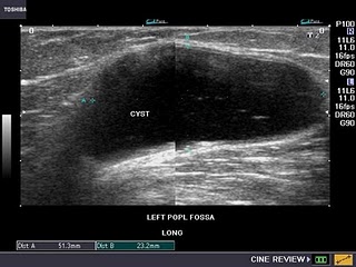
Many Americans are aware of the debate raging over the safety of CT scans performed. Issues regarding high radiation doses and appropriateness in the clinical setting have surfaced in recent months through out the media and in the blogosphere. The science behind radiation is clear: there is no such thing as a safe X-Ray. Medical professionals are taught in school that diagnostic X-Rays are to be used in the clinical setting when other, less hazardous forms of medical imaging will not give us the information we seek. Then why is it that when we go to the ED with an stomach pain we are sent to the CAT scanner instead of given an ultrasound? The answers are complex and troubling. In many cases the Physician trusts the result of the CT scan over the result of an ultrasound because they think the CT scan "Cannot miss anything". Many Physicians are under the impression that an ultrasound test is very operator dependent (True) and are loathe to trust the results for fear of being held liable if the test misses something. As a Sonographer, I agree that the results of the sonogram can be influenced by many factors including operator training and the skill of the interpretation, patient body habitus, and type of equipment used, but is CT really the gold standard? Should we really be subjecting our children and women of childbearing age to the risk of X-Rays without giving the matter much consideration? I would like to post part of an article I found on the web that discusses one of these issues. I welcome your feed back.

ScienceDaily (Mar. 8, 2010) — In a bold, eye-opening editorial in the March 2010 issue of the Journal of Ultrasound in Medicine, Harvard Professor, Beryl Benacerraf, MD, urges the medical community to use ultrasound instead of Computed Tomography (CT) as the first-line imaging test for better diagnosis capability in the evaluation of acute female pelvic and lower abdominal conditions.

"How have we evolved to ordering the most expensive imaging technique first for these patients, only to be followed frequently by a far less costly ultrasound examination to clarify the CT findings? Ultrasound is the established modality of choice to evaluate the female pelvis, so why do patients with pelvic masses or pain get a CT scan? In my opinion, doing a CT scan first for female patients with lower abdominal pain is dangerous and wasteful, a drain of much-needed health care dollars."
You can find the article here: http://www.sciencedaily.com/releases/2010/03/100302123122.htm?utm_source=feedburner&utm_medium=feed&utm_campaign=Feed%3A+sciencedaily+%28ScienceDaily%3A+Latest+Science+News%29

Thank you Science Daily.

Tuesday, March 9, 2010

First in Texas


I am pleased to announce I am the first Sonographer in the state of Texas to use the GE portable ultrasound machine. As you see, it is slightly larger than an I phone, and a wee bit heavier. My GE medical rep, Mr S. Potter brought the instrument over to my office for a test drive yesterday. First Impression: Nice images. Very clear echocardiogram. Nice frame rate, and color flow Doppler that was pretty darn good. No pulse or CW Doppler. Only one sector probe. Three presets: OB, ABD, and Echo. One issue: it got very warm after 10 minutes of scanning, and automatically put itself in an energy saving mode (Wonder if that is connected to the heating???) The energy saving mode reduced the frame rate to 3 hertz. All in all, a great product for a quick look see. More later. Peace....

Saturday, March 6, 2010

Contrast and Molecular Imaging with Ultrasound

An exciting application of ultrasound contrast technology has been recently presented which uses molecular interactions between gas filled contrast agents tagged with substances that bind to tumors. This is exciting because we may be able to spot cancers while they are very small, and treat them before they spread.

"ScienceDaily (Mar. 6, 2010) — An imaging technique combining ultrasound and specially modified contrast agents may allow researchers to noninvasive detect cancer and show its progression, according to research published in the March issue of The Journal of Nuclear Medicine (JNM). The technique enables researchers to visualize tumor activity at the molecular level." Please see the full post here: http://www.sciencedaily.com/releases/2010/03/100301102751.htm

Thanks Science Daily. I hope further research is fruitful in this area. This seems like it may be applicable to many types of cancer, and perhaps, other diseases.

Editor

Friday, January 8, 2010

ED Physicians Ultrasound Test Available

The American College of Emergency Physicians (ACEP) http://www.acep.org/ has published an on-line self test for emergency physicians to take when they are ready to document that they are proficient in the use of ultrasound in the practice of emergency medicine. This is the first test that I know of to address the issue of self credentialing. I took a few modules of the test myself (I passed) and found the test to be challenging, though I have been practicing ultrasound for many years as a Sonographer. The modules are of the multiple choice form with embedded videos that one may click on for clarification. One should read the question carefully before answering as the question itself can be confusing. I found at least two wrong answers listed as the correct response. For example: a patient is NOT routinely placed in the PRONE position to image the gall bladder. I am sure these issues will be ironed out. I do believe this is a great first step in beginning a standardized pathway for ED physicians to obtain self credentialing at the institutions where they are employed. Currently, there is no charge for taking the test, and results can be printed out. Here is the link: http://emsono.com/acep
Thanks Dr Morale

Tuesday, January 5, 2010

Pneumothorax by U/S?


Recently,methods have been published outlining a technique for identifying moderate to sever pneumothorax using high frequency ultrasound. This technique is time saving and rather simple to perform at bedside and has been embraced by many ED Physicians in the trauma department. It involves looking for the presence or absence of a sliding lung using intercostal technique. Below is a link to one paper discussing this exciting new application of diagnostic ultrasound. Happy New Year.

Article Title: Sonographic Detection of Pneumothorax by Radiology Residents as Part of Extended Focused Assessment With Sonography for Trauma
Authors: Olga R. Brook, MD, Nira Beck-Razi, MD, Subhi Abadi, MD, Janna Filatov, MD, Anat Ilivitzki, MD, Diana Litmanovich, MD and Diana Gaitini, MD
A recently published article in the Journal of Ultrasound in Medicine assessed the accuracy of sonographic pneumothorax detection by radiology residents as a part of extended focused assessment with sonography for trauma (eFAST).

A sonographic search for pneumothoraces was performed as part of a standard FAST examination by the on-call resident. Each lung field was scanned at the second to fourth anterior intercostal spaces and the sixth to eighth midaxillary line intercostal spaces. A normal pleural interface was identified by the presence of parietal-over-visceral pleural sliding –“lung sliding sign”. Absence of lung sliding and comet tail was presumed to be indicative of a pneumothorax.

The sonographic diagnosis was correlated with supine chest radiography and chest CT. A total of 169 patients were included in the study. Chest CT was considered the reference standard examination. CT chest identified 43 pneumothoraces (13%): 34 small and 9 moderate.

On chest radiography, 7 pneumothoraces (16%) were identified. Extended FAST identified 23 pneumothoraces (53%). Compared with CT, eFAST had sensitivity of 47%, specificity of 99%, a positive predictive value of 87%, and a negative predictive value of 93%.

All of the moderate pneumothoraces were identified by eFAST. Twenty small pneumothoraces missed by eFAST did not require chest tube placement during the hospitalization period.

The authors concluded that the eFAST exam for detection of pneumothoces is an accurate and efficient tool for detection of clinically significant pneumothoraces. However, small to moderate pneumothaces may be missed.

To read full abstract go to:
http://www.jultrasoundmed.org/cgi/content/abstract/28/6/749

Thank You Sonoworld for the link